Rethinking Joint Relocation to Enhance Patient Care
Do you want the best possible care for you and your family? Thanks to your support of the RAH Research Fund, people like John Au are able to ensure this high quality care is available.
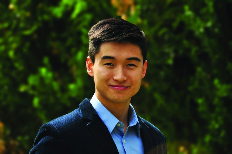
As a Medical Honours student from the University of Adelaide, John is taking part in an important project at the RAH investigating teaching methods on joint relocation for medical students, and looking at how they can be improved.
“Studies have shown the ability for a doctor to be able to relocate joints in patients is actually declining and the reason is historically there hasn’t been a standardised teaching method in medical school here in Australia. Joint relocations is an important skill that doctors should be able to do confidently and I’m excited to be able to play a role in giving them that confidence,” John said.
A joint is where two bones meet and dislocating a joint can cause a lot of pain and discomfort. Joint dislocation is quite common, and is mostly a result of age or a sports injury. It is an injury that needs prompt treatment as it can impact on nerves and muscle damage.
“In medical school you are taught theoretically, and as a medical student myself, I know that we are expected to know procedural techniques despite having never done them before, maybe only seeing it being done by a senior doctor,” John explained.
“This led to our project on the teaching methods of medical students where we conducted procedure skill workshops with students using cadavers, which are bodies that have been donated for medical science research. We wanted to see how effective using cadavers can be to teach medical techniques of joint relocation.
“We randomly divided 146 fourth year medical students studying at Adelaide University into five groups, each with different teaching methods. Those methods included instructional cards with photos showing how to do a joint relocation step by step and a skeletal model where they could move around and visualise how they can relocate the joint. The third group got a cadaver that had been dissected so the joint was dis-locatable and relocatable which allowed the students to practice going through and relocating joints, and to also get a feel for it. The last two groups received skeletal models, one focused on a patellar (kneecap) relocation and the fifth group looked at the hip and knee relocation joints.”
John explained that during this process each group received instructional cards as a baseline at the beginning of the teaching intervention, and at the end they received a survey with 13 questions.
“These questions were based on their understanding, familiarity of the techniques and also how comfortable and confident they feel in doing this procedure,” John explained.
“The results were as expected, each teaching method no matter what intervention saw an improvement and students felt more comfortable after the procedures. We found that using cadavers for medical research allows medical students to improve their hands on approach with procedures and allows them to feel confident and prepares them for real-life situations.
“Ultimately, results did show using cadavers made a huge difference for medical students and it was an extremely effective teaching method. Cadavers can also be used for learning other medical procedures, not just joint relocation. Some can also last up to 10 years for medical research, it is an incredible thing to donate your body for medical research.”
It’s also an incredible thing to donate funds to enable this medical research to happen! Your support through the RAH Research Fund enables this, thank you!